Affinity Purified Rabbit Polyclonal Antibody
| 货号 | 规格 | 价格 |
|---|---|---|
| P900003 | ||
| 20µL | ¥588.00 | |
| 50µL | ¥1080.00 | |
| 100µL | ¥1780.00 |


| Product Name | Anti-PDIA1 (PDI) Rabbit pAb |
|---|---|
| Description | Affinity Purified Rabbit Polyclonal Antibody |
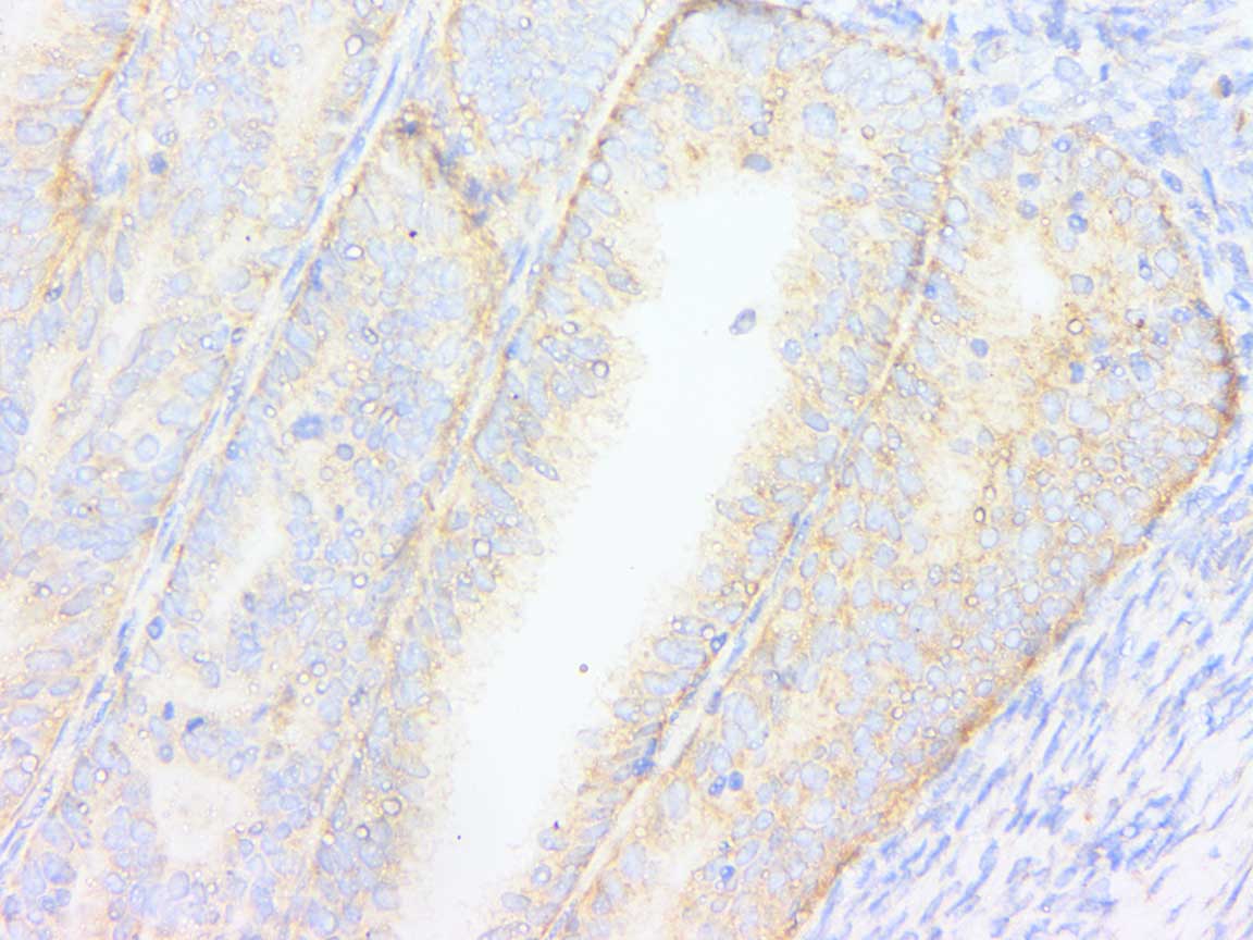
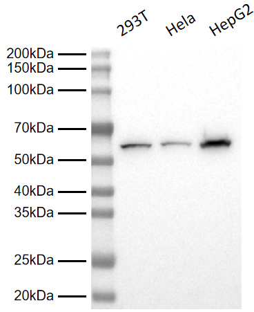
Application
|
WB, IHC-P, IF(Cell), ELISA |
| Dilution | WB 1:5,000; IHC-P 1:200~1:1,000; IF 1:50~1:100 |
| Reactivity | Human |
| Host | Rabbit |
| Clonality | Polyclonal |
| Isotype | IgG |
| Target / Specificity | This PDI antibody is generated from rabbits immunized with a BSA conjugated synthetic peptide between 488-502 amino acids from the C-terminal region of human PDI. |
| Format | Purified polyclonal antibody supplied in PBS with 0.01% (W/V) sodium azide and 50% glycerol, pH 7.3. This antibody is purified through a protein G column. |
| Gene ID | 5034 |
|---|---|
| Synonyms | P4HB,ERBA2L, PDI, PDIA1, PO4DB |
| Calculated MW | Calculated MW: 57 kDa; Observed MW: 57 kDa |
| Primary Accession | P07237 |
| Other Accession | NP_000909.2 |
| Antigen Region | 488-502aa |
| Storage | Shipped at 4℃. Upon delivery aliquot. Store at 4℃ short term (1~2 weeks). Store at -20℃ for 2 years. Avoid freeze / thaw cycles. |
| Precautions | Anti-PDIA1 (PDI) Rabbit pAb is for research use only and not for use in diagnostic or therapeutic procedures. |
| Background | This multifunctional protein catalyzes the formation, breakage and rearrangement of disulfide bonds. At the cell surface, seems to act as a reductase that cleaves disulfide bonds of proteins attached to the cell. May therefore cause structural modifications of exofacial proteins. Inside the cell, seems to form/rearrange disulfide bonds of nascent proteins. At high concentrations, functions as a chaperone that inhibits aggregation of misfolded proteins. At low concentrations, facilitates aggregation (anti-chaperone activity). May be involved with other chaperones in the structural modification of the TG precursor in hormone biogenesis. Also acts a structural subunit of various enzymes such as prolyl 4-hydroxylase and microsomal triacylglycerol transfer protein MTTP. |
|---|
| Cellular Location | Endoplasmic reticulum. Endoplasmic reticulum lumen. Melanosome. Cell membrane; Peripheral membrane protein. Note=Highly abundant. In some cell types, seems to be also secreted or associated with the plasma membrane, where it undergoes constant shedding and replacement from intracellular sources (Probable). Localizes near CD4-enriched regions on lymphoid cell surfaces (PubMed:11181151). Identified by mass spectrometry in melanosome fractions from stage I to stage IV (PubMed:10636893) Colocalizes with MTTP in the endoplasmic reticulum (PubMed:23475612) {ECO:0000269|PubMed:10636893, ECO:0000269|PubMed:11181151, ECO:0000269|PubMed:23475612, ECO:0000305}. |
|---|
| Tissue Location | Stromal cell of endometrium, parotid gland, body of pancreas, jejunal mucosa, ileal mucosa, islet of Langerhans, right lobe of liver, duodenum, endometrium epithelium, lower esophagus mucosa, adenohypophysis, type B pancreatic cell, minor salivary gland, left adrenal gland, left adrenal gland cortex, right adrenal gland. |
|---|

Copyright © 2023上海雅酶生物医药科技有限公司(Epizyme Biotech)
沪ICP备2021002422号